It is important to take the time to understand the anatomy of the human eye and how vision works. This knowledge will help you and your doctor better understand your symptoms.
How Vision Works
The eye is a small but complex organ. It is this organ that many people believe provide us with one of the most important senses, vision.
Vision occurs when light enters the eye through the pupil. Other structures, like the iris and cornea, help direct the appropriate amount of light towards the lens.
It helps to think of your eye lens working like a camera lens would. When you take a picture with your camera lens, it sends a message that produces a picture. Well, your eye lens bends incoming light onto your retina. Millions of specialized cells within the retina work together to transform the image, which is then processed by the brain.
Eye Anatomy
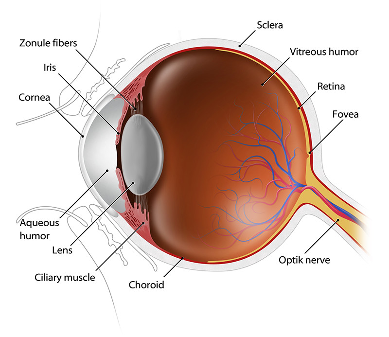
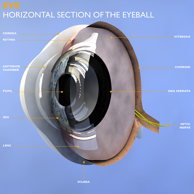
Cornea: the transparent circular part at the front of the eyeball. It refracts (bends) the light that enters the eye onto the lens. The cornea is extremely sensitive to pain
Pupil: the circular opening in the centre of the iris. The pupil is where the light passes into the lens.
Iris: This forms the colored part of your eye and regulates the amount of light that enters your eye.
Lens: a transparent structure that sits behind your pupil. The lens helps to refract incoming light and focus it onto the retina.
Choroid: a middle layer that sits between the retina and the sclera. It contains a pigment that absorbs excess light to prevent vision blur.
Ciliary muscle: this part of the eye connects the choroid to the iris.
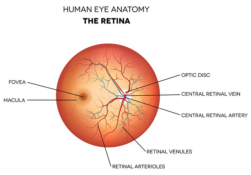
Retina: lines the interior of the eye and is composed of light sensitive cells called rods and cones. Rods are necessary for seeing in dim light, while cones function best in bright light.
Optic disc: identifies the start of the optic nerve. This is where the messages from the cone and rod cells leave the eye to reach the brain.
Optic nerve: transfers all the visual information to the brain.
Sclera: the white part of the eye.
